Operative hysteroscopy
A very conservative form of treatment of intrauterine disease
Operative Hysteroscopy is a very conservative form of treatment of intra cavitary disease. The uterus remains in place and hence can give rise to other diseases in the future.
There is one "Standard of care" in operative hysteroscopy: the hysteroscopic septum resection:
- No incision in the uterine fundus hence no Caesarean Section for mechanical reasons at the end of the pregnancy.
- Minimal theatre occupancy: a mean theatre time of 30 min including the actual theatre time of 8-15 minutes.
- Minimal technical needs: simple mechanical scissors or unipolar knive are sufficient.
- Day Hospital cases: all of the septum resections.
- Patients resume their daily activities fully within three days.
- High patient satisfaction.
- Exactly the same medium to long term results as compared with the classical surgery concerning pregnancy rates.
In the long term even better results as there are no intra- abdominal scars as compared to the 45%-85% scars in classical open abdomen surgery.
The "Golden" standard is the best possible care you can provide your patient with. The "Standard of Care" is the care you have to give your patients routinely, if you do not provide your patients with this care you are liable in court.
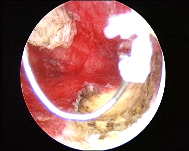
Hysteroscopic myoma resection is justified only when there is an active desire for further fertility or when the patient refuses a hysterectomy. The risk of intra-vasation of distention medium outweighs only a good indication. Intravasation is highly likely because relative large blood vessels have to be transsected. Only myomas that are imbedded up to 50 % in the muscle coat of the myometrium are an indication. Deeper lying myomas should be operated by experts in hysteroscopic surgery to minimize the risks for the patients. Fig. 3 gives a view of the bleeders in the normal myometrium, pink as compared to the whiter color of the myoma fibers, that we have to coagulate as we go along to avoid excessive intravasation of distention fluid. At the end we see the fundus and the implantation zone is often small as compared to the bulk of the myoma obstructing more or less the whole cavity fig. 4.
Hysteroscopic endometrial resection: this technique is in competition with the "global endometrial destruction" techniques. The advantages of the hysteroscopic technique is that it becomes possible to treat polyps and small myomas at the same time as the endometrium with better results in the long run, if amenorrhoea is the referal point, than the other techniques. The learning curve is a handicap but the main advantage is that there is tissue to give to the pathologist for evaluation. Small endometrial carcinomas have been described that were missed with currettage and diagnostic hysteroscopy but found in the chips at endometrial resection ( Colafranceschi M., Bettocchi S., Mencaglia L., Van Herendael B.J.. " Missed Hysteroscopic detection of uterine carcinoma before endometrial resection: report of three cases." Gynecol Oncol 1996;62:298-300.). Manifest abnormal looking endometrium ( as in Fig. 2 ) should therefor not be operated upon. A biospy has to be taken and a pathology report awaited before action is undertaken. The visual impression alone is not good enough. |
|